
Personal Protection from harmful agents within the cabinet
Product Protection to avoid contamination of the samples.
Environmental Protection from contaminants contained within the cabinet.

The stability chamber are used to study the impact of pre-specified circumstances on life-saving pharmaceuticals, biological samples, electronic components, and industrial parts, among other things. Before getting into the different types of stability chambers, let’s find out about a few important features of this product.
Six Sizes of Stability Chambers
Model Volume
IGL-SC200 200 Liters
IGL-SC324 324 Liters
IGL-SC450 450 Liters
IGL-SC600 600 Liters
IGL-SC800 800 Liters
IGL-SC1000 1000 Liters
IGene Labserve offers some of the best quality stability chambers, various optional changes can be made in this product over suggestions like:
- Stainless steel exterior (GMP)
- RS 485 interface
- PLC based HMI controller w/ USB interface
- 21 CFR part 11software
- Storage racks
- UV light
If there is anything you are looking to understand about stability chambers, then visit their official website at https://www.igenels.com or dial 09310696848 to speak to them directly.

A gel documentation system is mainly used in molecular biology labs with an aim for imaging and documenting nucleic acids and proteins that are suspended in polyacrylamide or agarose gels. These gels are stained with fluorophores like ethidium bromide or SBYR green. Most of us haven't come across gel documentation devices in real life as they are only used for scientific purposes. Simplistically, a gel doc comprises a light source, CCTV camera, transilluminator, and a dark room to protect the body from UV rays. For this reason, it is also known as a gel image system or a gel imager.
Gel documentation machines are widely used in genetic engineering laboratories. Be it colleges, protein-generating pharmaceutical industries, or genetic engineering research institutes. Moreover, gel documentation mechanisms are also used in clinical research labs and forensic laboratories.
Gel imaging is all about recording and identifying nucleic acid and labeled protein in diverse media like agarose, cellulose, and acrylamide. Microbiologists use this system for the rapid assessment of gels post electrophoresis and thereby, saving chemicals and time. It leads to visualization in 3-4 minutes. Gel documentation devices use stain-free technology that makes them pocket-friendly in every aspect.
There are various types of gel documentation systems based on the sample type and throughput. Also, these systems come in different configurations and specifications that come in handy for the users. So, let's explore the diverse types of documentation systems:
The prime advantages of using a gel doc system are listed below:
A gel documentation machine is necessary for any lab performing protein analysis. In most instances, these systems come in diverse specifications with a customization option based on the level of application. Hence, it's always advisable to buy the one streamlined to your laboratory's needs while at the same time focusing on the budget.


If you wish to get a brief understanding of the working and the use of the stability chamber, this blog will provide you with answers, read on to see if your stability chamber is meeting all these requirements.
What is the use of stability testing in the chamber?
Placing samples in environmentally controlled chambers to determine their quality and product value over a period of time under the influence of various environmental changes such as temperature difference, humidity, and light changes tests the performance of the sample. This data is useful in the research and development stage of any product before it is introduced to the market. The industries that greatly rely on this testing format are pharmaceutical manufacturers, medical device manufacturers those in the food manufacturing industry, dealing with life science samples, etc.
What is the reason for stability studies?
Stability studies are a must for certain industries such as those dealing with drug development. The stability testing results reveal the influence of physical, chemical, and microbiological reactions on a sample, the test reveals the efficacy of the sample and the integrity that the final product shows when the testing period is over. A stability study program ensures the commercialization of the product sample determining its shelf life and helping manufacturers take the next step to the product's development.
How is the stability testing done?
The sample is stored in environmentally controlled chambers as a part of the stability study program. The samples are kept for a time frame after which they are removed and tested to see if temperature, humidity, and light changes have impacted the physical, chemical, or microbiological characteristics of the sample.
What is the length of stability testing?
Stability testing is done according to the guidelines recommended by the ICH International Council for Harmonisation of Technical Requirements for Pharmaceuticals for Human Use (ICH). Broadly classifying stability in long-term testing extending for a minimum of 12 months at 25°C/60%RH, the long-term testing allows product degradation under conditions of recommended storage, intermediate testing is done at 30°C/65%RH. Accelerated testing is done for a minimum of 6 months at 40°C/75%RH. The product goes through a short-term testing period.
Retain testing is done on products that will be marketed from a batch of finished products defining market authorization, labels and packaging, and pharmacy vigilance report.
Thermal cycling testing mimics conditions like marketplace storage and the reaction it will have when it is transported to the market, on loading and offloading, etc.
How is accelerated testing done?
Storage for stability testing follows 40°C +/- 2 degrees and Relative Humidity (RH) of 75% +/- 5% according to the ICH guidelines. This testing is based on the final formulation of the established manufacturing process. The test determines shelf life and storage conditions in real-time studies.
How is long terms testing done?
Also called the room temperature stability storage, this testing condition follows storage parameters measuring according to ICH guidelines. This is a forced degradation study that provides more knowledge on the drug development process, the degradation chemistry, of drug substances and products. The finding is an authority that builds the foundation of drug formulation and packaging development. The ICH does not state any guidelines for forced degradation, but 5-20% is the recommended degradation limit.
What is photostability testing?
This testing is a must for ensuring quality especially for manufacturing pharmaceutical drugs. If the drug is not tested adequately it will result in additional cost failure of the drug and loss of revenue. The selected drugs go through single batch testing, if the batch cites variations then the studies are repeated.
Reference samples are used for analytical testing of fully finished products.
Retention samples are used for identification purposes that include presentation, packaging, labeling, patient information leaflet, batch number, expiry date.
Both reference and retention samples are considered before a finished product is made ready for the market.
Conclusion
There is more to the stability chamber that those using it must know. You can go into greater details on the product from our website https://www.igenels.com/ and also purchase your stability testing device from us for the best results.

What is PCR?
A
polymerase chain reaction workstation is enclosed on all three sides.
Amplifying DNA and RNA fragments into millions of copies this
workstation is used in life science laboratories. The equipment prevents
cross-contamination.
What is the working of a PCR machine?
What more can you find from iGeneLabserve PCR workstation?
LCD air velocity display,
lamp on/off time status display,
Germicide UV lamp sterilizing the workspace before starting work
Add extra electric sockets
Add Magnehelic gauge if required.
If you purchase your PCR workstation
from IgeneLabServe, check the details in the product manual to make
your PCR workstation more customized. Look out for the added features
that you require. Visit the website https://www.igenels.com/. Call 1800
5720 603 for details.

Gel Documentation System measures
and records labeled protein and nucleic acid; different media like
acrylamide, cellulose, or agarose are used for the diagnosis. The
equipment aids in quick evaluation after electrophoresis which proves to
be economical in terms of chemical and time. You can see the results in
less than five minutes through stain-free technology. This testing
equipment is used in research labs that help you to visualize
photo-documented nucleic acid separated samples taking place after
electrophoresis, show microbial colonies count and protein separation on
western blots, and thin layer chromatography (TLC) mixture
identification.
What is the basic working of a Gel Imager?
The gel imager comes with an ultraviolet (UV)/ visible (blue or white) transilluminator. The light source is blocked from penetrating with a hood and inside the equipment; a high-resolution camera captures the image. The procedure involves samples mixed with the agarose gels placed on a Petri dish or TLC films, placed on the transilluminator surface. The door closes down and prevents the UV light from penetrating outside harming the user. Once the lamp is powered the photo documenting through CCD and CMOS cameras takes place. The images can be retrieved and exported on Microsoft Excel.
Labs that use the gel images
Used in molecular biology labs for genomics, proteomics, the imaging system identifies PCR segments with DNA quantification, bacterial cell culture, protein separation, and environmental sample testing. Emitting visible light or Ultra Violet light for visualizing chemiluminescent samples or bacterial sample count, the UV light should match the absorption spectrum of the fluorescent dye used at the time of gel separation through a blotting and thin chromatography process. For colony counting at its optimal amount, the blue light emitted at 470 nanometers and staining samples through commercial dye commonly use SYBR Green, SYBR Gold, or SYBR Safe. Visualize, UV 254 nanometer light at its optimal amount to identify DNA cross-linking, for short exposure gels, stained with Ethidium Bromide 302 nanometer light is best suited and for gel band cutting, 365 nanometer light suits perfectly.
Gel Documentation System Application and uses
B1 - horizontal minigel system
For identifying DNA and RNA samples for analysis, cloning, and sequencing, samples of DNA and RNA (Necluic acids)are loaded on agarose or acrylamide gel. Exposing them to the electric field, the negatively charged samples travel towards the positive electrode. The smaller DNA and RNA fragments travel faster than the longer ones, enabling fragment identification aided by the fluorescent dyes. Once identified, the samples are separated for purification.
B2 - horizontal minigel system
The protein sample is immersed in Tris-Glycene or Tris-Acetate– Tris-Glycene or Tris-Acetate which is commercially available and is effective in purification and separation. The protein fragments travel at different speeds through the gel metrics. Mass spectrometry, sample denaturing, and blotting use the protein gel.
B3 Colony counting
Microbial colony-forming units’ optimal growth is determined within the petri dish that has cell media loaded in it.
B4 Thin layer Chromatography
Quantification and qualification of a mass of substances include lipids, carbohydrates, fatty acids, and pesticides.
B5 Southern and Western Blot Gel
Separates proteins and isolates them for purification of active gels and SDS-PAGE gels.
Southern Blotting Gels
Separation of DNA fragments from blood and tissue samples restricting enzyme digestion through polyacrylamide (PAGE) gels and sodium dodecyl sulfate (SDS) gels combining urea.
Conclusion
A necessity for any lab that demands protein analysis, the imaging system comes in a range of specifications. However, you can customize the level of application if you order your gel imager from iGene Labserve. Visit our website and get in touch for details https://www.igenels.com/. You can also explore our gallery of scientific equipment to expand your lab assets.

One of the most significant and advanced molecular biology instruments, the PCR Machine has created a new chapter by revolutionizing DNA study. Kary B. Mullis was awarded a Nobel Prize for his invaluable discovery and contribution to Chemistry.
After amplification from the machine, the DNA is used in various lab procedures like human genomic projects (HGP), laboratory and clinical techniques that include DNA fingerprint, bacteria detection, and genetic disorder diagnosis.
How is the amplification of DNA segment done in PCR?
After heating the sample to denature DNA or separate the two strands of DNA into a single strand. With an enzyme, "Taq polymerase" synthesizes building new DNA strands from the original strands that are termed templates. With this process of duplication from the templates, each molecule contains an old and new DNS strand. Each strand creates two new copies and the process goes on. This cycle is repeated 30-40 times of synthesized and denatured DNA producing more than a billion copies from the original segment. The machine works in an automated format and is capable of completing the procedure in a few hours; the thermocycler machine can alter the temperature automatically as it is programmed to denaturing and synthesize every minute.
What are the requirements that your system must have?
A single PCR is unable to accommodate the requirements of the research community. For genomic application, an optimally configured rtPCR device is good for most laboratories. The designs are varied with Peltier elements accommodating tubes and microtiter plates. For countering the “edge effects” of achieving different temperatures, different approaches are used in overcoming the problem. In some cases independent thermal electric modules monitor each sample in different phases; in some machines, a carousel system spins the sample in temperature-controlled cabins maintaining equalized thermal stability.
Some companies work on closed systems using specific reagents or reaction vessels/ kits. This makes the entire assembly expensive and with limited flexibility. The reaction speed of the machine matters, some machines have robust throughput allowing standard rtPCR reaction that can be completed in half an hour to a little less than an hour producing 384 well plates.
Conclusion
Standalone PCR systems computer-controlled with user interface come with data storage facilities and network options. A careful study of what you require from your PCR machine is necessary before you select the right instrument. Meeting your specific laboratory requirements and pricing considerations, iGene Labserve brings the most convenient closed cabinet-fitted HEPA filtered airflow systems that maintain minimum turbulence with maximum output. Visit https://www.igenels.com/ for carrying out specialized work in a bacteria-free environment.

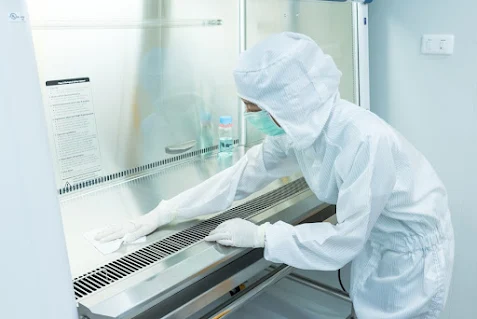
Biosafety Cabinet (BSC) is a laboratory-controlled lab-engineered enclosure that protects samples, lab workers, and the laboratory environment against exposure and biohazards, splashes, and aerosols. The chances of material splashes and aerosols arise from material handling and manipulation of highly communicable agents. It is necessary to periodically check the efficiency of safety cabinets to verify the airflow, HEPA integrity, and how well the instrument is being able to protect the sample stored inside the chamber. All these factors depend on the cabinet in use.
The Biological safety cabinet comes in three classes. Class I cabinets, Class II cabinets, and Class III cabinets. Let's go through a quick review of what each entails.
Class I cabinet
This is a simple cabinet that provides personnel and environmental safety. However, in this type of cabinet, the product is subject to contamination. The airflow suctioned inside the cabinet is discharged through the HEPA filters.
Class II cabinet
This ventilated cabinet provides both personnel, environmental, and product safety. Found in research and clinical labs this is ideal for labs that deal with infectious agents. This cabinet is further divided into four three risk groups. A1, A2, B1, B2 are the different types of class II cabinets that are differentiated by the ratio of their exhaust and air recirculation.
Class III Cabinets
This cabinet provides the highest level of safety and is used extensively for protecting the most transmissible agents. These cabinets are used in Level 3 and 4 laboratories. These are completely enclosed cabinets that are leak proof, with HEPA filtered air. The operator's access is provided using glove boxes that enable work in the enclosed workspace. Dedicated exhaust system exterior maintains an interior negative pressure.
Conclusion
Providing the most technologically advanced Biosafety Cabinet
iGenge labserve brings the most advanced laboratory equipment that
increases lab experiment success and opens scope to major and delicate
investigations in the field of bioscience. To explore our product
brochure visit https://www.igenels.com/
Setting up a new laboratory involves more than purchasing instruments and arranging them on benches. The equipment needs to match the labora...


